 We use our eyes constantly to see the world around us, but the workings of that small organ are sometimes mysterious. Though only about an inch in diameter, a healthy adult eye consists of a number of key parts that work together along with the brain to bring the outside world into our perception. What we think of as seeing – the representation and interpretation of images – actually takes place in the brain, but the eye is responsible for gathering all of this sensory data and aggregating it for transmission to the brain. For more interesting articles about eye health click here.
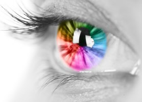
1 – The Cornea and Pupil
Beginning at the front of the eye, the first part that you will encounter is the cornea. The cornea is a layer of clear tissue that sits in front of the pupil and the iris and allows light to enter the eye, as though through a window. The cornea lets light through indiscriminately, while the pupil, the dark spot at the center of your eye, expands and contracts to determine how much light is absorbed. In a bright room, your pupil contracts to limit the amount of light entering the eye, while in the dark the pupil expands to absorb as much light as possible.
2 – How the Lens Works
After light passes through the cornea and the pupil, it passes through the lens of the eye. The lens of the eye performs the critical function of focusing the light that enters the eye. There are tiny muscles that surround the lens, allowing it to shift and deform and focus light at different distances. At a close range, the lens is drawn flat and thin, while at greater distances the lens is compressed to be thicker. In a healthy eye, the lens can delicately change shape to focus the eye on objects at a wide range of distances, while individuals who need glasses for nearsighted or farsightedness are less able to focus the lens of the eye.
3 – On to the Retina
When light passes through the lens, it then travels through the vitreous fluid. The vitreous fluid fills the majority of the eye and has the consistency of gelatin. On the other side of the vitreous fluid is the retina. The retina lies along the back of the eye and is the part of the eye that is sensitive to light. The retina is made up of two different types of cells: rods and cones. Rods are responsible for low-light vision and provide monochromatic perception, while cones are mainly used for color vision and higher detail vision. Cones come in three varieties: red, blue, and green.
4 – From the Retina to the Brain
The retina has several components to it. While the top layer is primarily made up of rods, there is a layer right below the retina called the fovea where cones are congregated. Another important part of the retina is the macula. The macula is densely populated with cones and is the main part of the retina responsible for detail vision. Visual information congregates at the macula where it is then transmitted to the optic nerve, which carries the visual information from the eye to the brain where it is processed and interpreted.